


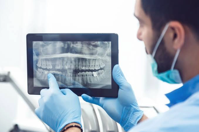
There is no one shot that is best for every patient. Sometimes a small image of one or several teeth is sufficient, while an orthopan or 3D CBCT image is required for an examination of both jaws, implant planning or a more complex surgical intervention. The choice should be made by the dentist after a discussion, examination and insight into previous images.
The most important in a nutshell: imaging is quick and painless, special preparation is generally not required, and the radiation dose with modern dental devices is small. However, X-rays are not done automatically or on a calendar, but only when they may change the diagnosis, treatment plan or follow-up results.
What is a dental scan and what can the dentist see on the scan?
The term “dental imaging” usually refers to dental radiography, i.e. the use of X-rays to obtain an image of the teeth, jaws and surrounding structures. Depending on the type of image, the dentist can assess:
- caries that is not visible during clinical examination, especially between teeth and under existing fillings;
- shape, number and length of roots and canals;
- inflammation or change around the tip of the root;
- the level and quality of the bone around the teeth;
- position of unerupted, supernumerary and impacted teeth;
- the relationship of teeth to nerves, sinuses and adjacent anatomical structures;
- condition after root canal treatment, tooth extraction, installation of implants or other interventions;
- jaw relations and tooth position important for orthodontic therapy.
The image is part of the diagnosis, not a diagnosis in itself. The findings are interpreted together with the patient’s complaints, clinical examination, previous scans and other relevant data.
Types of dental radiographs
Small tooth scan – retroalveolar or periapical scan
A small, intraoral image shows one tooth or several adjacent teeth, from the crown to the tip of the root and the surrounding bone. It is most often used for toothache, suspected inflammation around the root, root canal treatment, assessment of deep caries, control after endodontic therapy and checking of the condition of the tooth’s supporting tissue.
Its advantage is a very detailed representation of a small region with a low dose of radiation. The limitation is that it does not provide an overview of the entire jaw.
Bitewing recording
Bitewing shows the crowns of the upper and lower side teeth in the same shot, as well as the area of bone between them. It is particularly useful for the early detection of caries between teeth, under the edges of fillings and crowns, as well as for assessing the level of alveolar bone in gum disease.
Occlusal image
An occlusal scan shows a larger area of the upper or lower jaw. Today, it is used less often, but it can be useful for determining the position of an unerupted or supernumerary tooth, evaluating an injury, major changes or a foreign body, especially when no other technique is available or suitable.
Orthopan – panoramic image of teeth and jaws
Orthopan, or orthopantomogram, is a two-dimensional panoramic image of both jaws, all teeth, jaw joints and part of the maxillary sinuses. It is often requested before the extraction of wisdom teeth, at the beginning of orthodontic therapy, when assessing the general condition of the teeth and bones, as well as for the initial planning of larger dental works.
Orthopan provides a good overview of a wide region, but due to magnification, overlap and two-dimensional display, it is not sufficient for every question. It should not be seen as a substitute for a small image when a very precise representation of a single tooth is required, nor as a substitute for 3D CBCT when bone width and spatial relationships are important.
Teleroentgen – cephalometric recording
A telex-ray usually shows the head in profile, and sometimes from the front. The orthodontist uses it to measure the relationship of the teeth, jaws and soft tissues of the face, analyze growth and plan therapy with fixed prostheses or other orthodontic appliances.
3D dental imaging – CBCT
CBCT, i.e. cone beam computed tomography, provides a three-dimensional image of the teeth, roots, jawbone and surrounding structures. The image can be viewed in multiple planes and sections, so the dentist receives information that cannot be reliably seen on a 2D image.
CBCT is most commonly used for:
- planning the installation of dental implants and assessing the amount of bone;
- determining the relationship of the wisdom tooth to the mandibular nerve;
- lokalizaciju neizniklih i prekobrojnih zuba;
- complex endodontic cases, additional canals, resorptions and suspicion of a root crack;
- assessment of cysts, other bone changes and injuries;
- selected jaw joint and sinus problems;
- planning guided implantological and other oral surgical therapy.
CBCT is not routinely performed on every patient. Compared to classic dental scans, it usually carries a higher dose of radiation, so it is chosen when a 2D scan cannot provide the data needed for a safe diagnosis or treatment plan. The size of the imaged field should be limited to the region of interest.
Are 3D CBCT and classic CT the same?
They are not. Both examinations use X-rays and provide cross-sections, but dental CBCT is adapted to imaging the teeth and bony structures of the face. In appropriate dental indications, it usually provides a very detailed image of the bone at a lower dose than a classic multi-slice medical CT scan. Classic CT is more suitable when evaluation of soft tissues or a wider medical region is important. The choice of examination depends on the diagnostic question.
Orthopan or 3D recording – what’s the difference?
| Characteristic | Orthopan | 3D CBCT |
| View / Visualization | 2D panoramic image of both jaws | 3D view and multiplanar cross-sections |
| Most Common Use | General exam, wisdom teeth, initial orthodontic and surgical assessment | Implants, complex surgery and endodontics, precise spatial relationships |
| Bone Width Display | Cannot reliably measure it | Enables three-dimensional assessment |
| Radiation Dose | Lower | Usually higher and depends on field of view size and protocol |
For the installation of the implant, orthopan can be useful as an initial examination, but the final plan is usually made on the basis of a 3D image, because it shows the width and height of the bone and the relation of the planned position of the implant to the nerves and sinuses.
A digital impression is not an X-ray
Patients sometimes confuse CBCT with an intraoral 3D scan. The intraoral scanner optically captures the surface of the teeth and gums and creates a digital model that can replace a classic impression. It doesn’t use ionizing radiation, but it doesn’t show the roots, the inside of the bone, the nerves, or the inflammation around the root tip. That’s why digital impression and X-ray have different roles and often complement each other.
When can a dentist ask for dental imaging?
An image may be needed when there is pain, swelling, injury, fistula, sensitivity when biting, suspicion of hidden caries or infection, as well as before root canal treatment, tooth extraction, prosthetic work, implantology or orthodontic therapy. It is also used to control healing or the results of treatment when such control may affect the further procedure.
The fact that the tooth does not hurt does not mean that the recording is unnecessary, but the absence of a recent recording is not in itself a reason to automatically take a new one. Contemporary recommendations emphasize individual assessment based on health and dental history, clinical examination, and existing imaging.
How often should teeth be photographed?
There is no rule that everyone has to have orthodontics done once a year or that the teeth are photographed at every check-up. Frequency depends on age, symptoms, risk of caries and periodontitis, previous treatment, planned therapy and quality of previous scans.
Save the previous images and show them to the new dentist. If they are of sufficient quality and still correspond to the current clinical question, they can prevent unnecessary repetition of imaging. The recording does not have a fixed “expiry date”: sometimes it is useful even after several years, while after an injury, a new complaint or a change in the therapeutic plan, a new recording may be needed very quickly.
What is the radiation dose during dental imaging?
The dose depends on the device, the setting, the size of the patient, the resolution and the size of the imaged field. The International Atomic Energy Agency lists the following typical effective doses:
| Type of dental scan | Typical effective dose |
| Intraoral scan | about 1–8 μSv |
| Orthopan | about 4–30 μSv |
| Teleroentgen | about 2–3 μSv |
| CBCT – small or medium field | medial about 50 μSv or less |
| CBCT – large field | medial about 100 μSv |
The values are approximate and do not represent the dose of each specific device. The effective dose serves to compare different procedures and cannot alone predict the individual risk of the patient.
For the protection of the patient, the most important thing is that the recording is justified, that the smallest field that corresponds to the diagnostic question is used, that the exposure is adapted to the patient and that the correct positioning reduces the possibility of repeating the recording.
Are lead apron and thyroid shield mandatory?
Pacijenti često očekuju olovnu kecelju ili kragnu, ali su savremene stručne preporuke promenjene. Američko stomatološko udruženje i Američka akademija za oralnu i maksilofacijalnu radiologiju više ne preporučuju njihovu rutinsku upotrebu pri dentalnom snimanju. Štitnik može da uđe u primarni snop, zakloni deo slike i dovede do ponavljanja snimka, dok su savremeni uređaji, ograničavanje polja i pravilna tehnika efikasnije mere zaštite.
Practices may vary depending on local regulations, devices and center protocols. The patient should follow the instructions of the trained staff, and he can clarify any doubt before the recording.
Dental imaging during pregnancy
Be sure to tell the dentist and X-ray center staff if you are pregnant or there is a possibility of pregnancy. Pregnancy, however, is not an automatic ban for any dental imaging. If the scan is not urgent and will not change current therapy, it can be postponed. When it is important to diagnose and safely treat an infection, severe pain, injury or other problem, it can be done with proper justification and an optimized protocol.
Since during dental imaging the pelvis is not in the direct beam, the dose received by the fetus is very small. The IAEA estimates that for dental examinations, including CBCT, the fetal dose ranges from approximately 0.009 to 7.97 μSv and that the risk of a few μSv is extremely small. If you took the scan before you found out you were pregnant, that in itself is not a reason to panic; inform the dentist and gynecologist for individual advice.
Dental imaging during breastfeeding
X-rays do not stay in the body or pass into breast milk, so after a standard dental imaging or CBCT examination without a contrast medium, it is not necessary to stop breastfeeding or to express and throw away the milk.
Dental imaging in children
A dental scan can be very important in a child because of cavities between the teeth, injury, monitoring of eruption, unerupted teeth or orthodontic problem. Since children are more sensitive to ionizing radiation, imaging should be individually justified, with a field and exposure adapted to the age and build of the child. CBCT is used in children only when a simpler method does not provide the necessary information.
What does the recording look like and how do you prepare?
- Bring a referral or write down clearly what type of image and what region the dentist was looking for.
- Bring previous scans if you have them, especially if following the same change or therapy.
- Food and drink are generally not limited; a standard dental scan does not require you to be on an empty stomach.
- Before orthopan, telex-ray or CBCT, earrings, piercings, glasses, chains, hairpins, mobile prostheses and other metal objects in the head and neck area are removed.
- During the exposure, you should remain still and follow the instructions of the technician. In panoramic and 3D recording, the camera rotates around the head, but the procedure is painless.
The exposure itself takes a short time, while with preparation, positioning and processing, the entire arrival usually takes a few minutes. If you have difficulty standing, cannot hold your head still for a long time, have a strong gag reflex or are afraid of closed spaces, tell the staff before the recording.
How is a dental image taken?
Digital 2D images can be obtained via e-mail, link, disk, USB or as a print, depending on the center. CBCT is most often delivered in DICOM format, sometimes with a viewing program, as a plain screen photograph does not contain all sections and data.
Save the original digital file and forward it to the dentist. For implant planning, it is important to submit a complete CBCT data set, not just a few printed images. Ask the center if the price includes a professional reading and a written report, as practice varies.
Where can dental imaging be done in Belgrade?
Below is an informative list of centers and surgeries in different parts of Belgrade. The list is not a ranking list or a recommendation of a particular service provider. Before arriving, check whether the center performs exactly the type of imaging that the dentist requested, whether an appointment is required, how the imaging is delivered and what the current working hours are. Contact information was verified on publicly available pages on July 20, 2026.
| Center / Clinic | Address | Phone | Website |
| Dental Scan | Venizelosova 3, Stari grad | 063 841 15 60 | dentalscan3d.rs |
| Studio za snimanje zuba | Takovska 58, Beograd | 011 69 00 635 064 11 00 120 | studiozasnimanjezuba.rs |
| Dental Diagnostic | Kraljice Natalije 36, Beograd | 011 26 56 036 065 265 60 36 | ortopanbeograd.rs |
| Rendgen Centar Beograd | Luke Ćelovića Trebinjca 34, Beograd | 011 7348 455 | rendgencentar.rs |
| C Dental Center | Cvijićeva 130, Beograd | 011 275 28 49 063 802 69 07 | cdentalcenter.rs |
| Bulevar 3D – Zvezdara | Jedrenska 4, Đeram pijaca | 011 403 77 51 062 288 833 | snimanjezuba3d.rs |
| ROorto 3D Digital | Bokeljska 1 (Bulevar oslobođenja 63a), Autokomanda | 011 244 10 21 069 244 10 21 | snimanjezuba.co.rs |
| 3Dent | Ustanička 170V, Voždovac | 011 454 4311 064 9999 489 | 3dent.rs |
| Bulevar 3D – Novi Beograd | Palmira Toljatija 5, lokal 105/L, TC Stari Merkator | 011 411 44 83 060 411 44 89 | snimanjezuba3d.rs |
| Belville Orto | Jurija Gagarina 14N, lokal 2, Novi Beograd | 011 6300 966 | belvilleorto.com |
| Ortopan Piramida | Jurija Gagarina 151G, Novi Beograd | 011 2280 329 065 257 1965 | 3dortopanpiramida.rs |
| Rendgen Centar Petković | Bulevar Mihajla Pupina 21/2, Novi Beograd | 069 212 1573 | snimanjezubapetkovic.rs |
| Dental Centar Luka Orto | Bežanijska 38, Zemun | 011 2610 201 060 5500 063 | snimanjezuba.com |
| Magic Dent | 22. oktobra 13, Zemun | 011 260 1638 065 260 1638 | magicdent.co.rs |


